veterinary dental x ray positioning pdf
Proper veterinary dental X-ray positioning is fundamental for accurate diagnosis and treatment planning․ It involves precise alignment of the X-ray beam and film to capture clear images of dental structures, ensuring optimal patient care and minimizing retakes․
Overview of Veterinary Dental Radiography
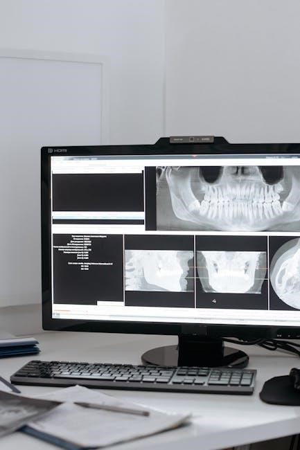
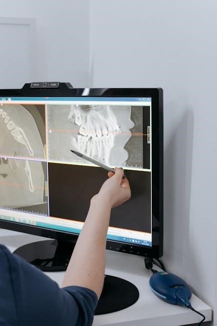
Veterinary dental radiography is a critical diagnostic tool for evaluating dental health in animals․ It provides detailed images of teeth, surrounding bone, and soft tissues, revealing abnormalities not visible during a physical exam․ Radiographs help identify fractures, resorption, periodontal disease, and other pathologies․ Intraoral and extraoral techniques are used, with intraoral being more common for detailed tooth views․ The bisecting angle technique is often employed to minimize distortion․ Proper positioning ensures accurate representation of structures, aiding in precise diagnosis and treatment planning․ This imaging modality is essential for veterinarians to assess dental conditions effectively, ensuring optimal patient care and outcomes․
Importance of Proper Positioning in Dental X-Ray
Proper positioning in dental X-ray is vital for obtaining accurate, undistorted images of dental structures․ Incorrect positioning can lead to elongation or foreshortening of teeth, making diagnosis challenging․ By aligning the X-ray beam perpendicular to the bisecting angle, images reflect the true size and shape of structures, ensuring precise diagnostic capabilities․ Proper positioning minimizes radiation exposure and reduces the need for retakes, improving efficiency and patient safety․ Accurate images enable veterinarians to identify hidden pathologies, such as root fractures or periodontal disease, which are critical for effective treatment planning․ Thus, precise positioning is essential for delivering high-quality care and achieving optimal outcomes in veterinary dentistry․
Basic Principles of Dental X-Ray Positioning
The core principle involves aligning the X-ray beam perpendicular to the bisecting angle of the tooth and film․ This ensures accurate, undistorted images for precise diagnosis and treatment planning․
The Bisecting Angle Technique
The bisecting angle technique is a cornerstone in dental radiography, ensuring accurate imaging by dividing the angle between the tooth’s long axis and the film․ Aligning the X-ray beam perpendicular to this bisecting angle minimizes distortion, providing clear, proportional images․ Proper execution requires precise positioning, with the beam angled to avoid foreshortening or elongation, ensuring the image reflects the actual size and structure․ This method is particularly crucial in capturing the apices of teeth for thorough evaluation․ By adhering to this technique, practitioners can consistently produce high-quality radiographs, essential for diagnosing dental issues in veterinary patients effectively․
Aligning the X-Ray Beam Perpendicular to the Bisecting Angle
Aligning the X-ray beam perpendicular to the bisecting angle ensures accurate and undistorted images of dental structures․ This technique involves positioning the beam so it forms a 90-degree angle with the bisecting line, which lies between the tooth’s long axis and the film․ Proper alignment minimizes foreshortening and elongation, providing a true representation of tooth length and surrounding bone․ The beam should be directed at the midpoint of the tooth, ensuring even illumination and clear visualization of the apices․ This method is critical for identifying pathological changes, such as root resorption or periapical lesions, and ensures diagnostic-quality radiographs․ By adhering to this principle, veterinarians can make precise diagnoses and develop effective treatment plans for dental conditions in animals․
Positioning for Specific Dental Structures
Proper positioning for maxillary and mandibular teeth ensures clear visualization of dental anatomy․ Techniques involve aligning the X-ray beam to capture accurate images of roots and surrounding bone structures․
Positioning for Maxillary Teeth
Positioning for maxillary teeth requires precise alignment to capture clear images of the upper jaw․ The X-ray beam should be angled to avoid distortion, ensuring accurate representation of tooth roots and surrounding bone․ For maxillary molars, the beam is often directed at a 90-degree angle to the bisecting angle, while premolars may require a slightly different approach․ Proper positioning involves placing the sensor parallel to the tooth row and adjusting the beam to perpendicular alignment․ This technique minimizes overlap and ensures detailed visualization of anatomical structures․ Guides like the Easy Guide to Dental X-ray Positioning provide specific angles and adjustments for optimal results․ Correct positioning is crucial for diagnosing pathologies and planning treatments effectively in veterinary dentistry․
Positioning for Mandibular Teeth
Positioning for mandibular teeth involves specific techniques to ensure clear visualization of the lower jaw structures․ The X-ray beam should be aligned perpendicular to the bisecting angle, minimizing distortion and overlap․ For mandibular molars and premolars, a lateral view is often used, with the film placed parallel to the tooth row․ The beam is typically angled at 90 degrees to the bisecting angle to capture accurate root structures․ For mandibular incisors and canines, a slightly different approach is required, with the beam aligned to avoid superimposition of surrounding bone․ Proper positioning ensures detailed images of the mandibular teeth and their surrounding anatomy, aiding in the diagnosis of pathologies such as root fractures or periodontal disease․ Referencing guides like the Easy Guide to Dental X-ray Positioning can help achieve optimal results for canine and feline patients․
Common Errors in Dental X-Ray Positioning
Common errors include improper beam alignment, insufficient perpendicularity, and incorrect angulation, leading to image distortion and magnification issues in veterinary radiography․
Identifying and Correcting Distortion in Images
Distortions in dental radiographs occur due to improper angulation or positioning, leading to elongation or foreshortening of tooth structures․ Identifying these issues requires careful analysis of the image․ For instance, if a tooth appears longer than its actual size, it indicates that the X-ray beam was angled too steeply․ Conversely, a shorter appearance suggests the beam was angled too shallowly․ Correcting these distortions involves adjusting the beam’s angle to ensure it is perpendicular to the bisecting angle of the tooth and the film․ Proper positioning guides, such as the bisecting angle technique, help minimize distortion, ensuring accurate representation of dental anatomy․ Regular calibration of equipment and technician training are essential for producing high-quality, undistorted images in veterinary dental radiography․
Advanced Techniques in Veterinary Dental Radiography
Oblique Views for Complex Dental Structures
Oblique views in veterinary dental radiography capture intricate tooth details by angling the X-ray beam, reducing overlap and enhancing visibility of complex structures like maxillary cheek teeth․
Oblique views in veterinary dental radiography are essential for evaluating complex structures, such as maxillary cheek teeth and mandibular teeth, by angling the X-ray beam 20 degrees․
This technique reduces overlap and enhances visibility of intricate details, aiding in diagnosing fractures, periodontal disease, or root abnormalities․
Proper positioning involves rotating the mandible or maxilla to align the beam with the area of interest, ensuring clear images for accurate diagnosis․
Oblique views are particularly useful for assessing teeth that are difficult to visualize in standard lateral or dorsal-ventral projections․
By adjusting the beam angle, veterinarians can obtain detailed images of specific dental structures, improving treatment planning and patient outcomes․
Regular practice and adherence to positioning guidelines are critical for mastering this advanced technique in veterinary dental radiography․